¿Puede un Absceso Profundo de Cuello, Llevarte a la MUERTE? | ¿Can a deep neck abscess lead to DEATH? [ESP]-[ENG]

Hola que tal mis queridos amigos de la #colmena, una vez más vengo por acá a traerles material innovador, fresco, el cual surge de una preocupación personal, dada la alta tasa de incidencias que me llegan a diario en la consulta de otorrinolaringología, en el cual, gracias a la mal higiene bucal, muchas vidas se encuentran comprometidas, y lastimosamente, un 20% de estos pacientes, no cuentan para contarlo, es así como les dejo la información, de que se trata el Absceso Profundo de Cuello.
Hello my dear friends of the #hive, once again I come here to bring you innovative, fresh material, which arises from a personal concern, given the high rate of incidences that come to me daily in otolaryngology consultation, in which, thanks to poor oral hygiene, many lives are compromised, and unfortunately, 20% of these patients, do not count to count it, so I leave the information, what is the Deep Neck Abscess.

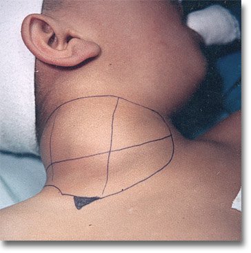
Son procesos patológicos que se desarrollan en un conjunto de espacios anatómicos, limitados por fascias y planos aponeuróticos, comprendidos entre la base del cráneo y una línea horizontal a nivel clavicular. Las infecciones de cualquier punto de la Vía Aerodigestiva Superior (VAS) se pueden diseminar a esos espacios de forma fácil y rápida, ocasionando graves complicaciones como mediastinitis, septicemia o fascitis necrotizante. Pueden comprometer la vía área. En ocasiones es difícil encontrar su origen, ya que la fuente primaria de la infección puede precederla en semanas.
These are pathological processes that develop in a set of anatomical spaces, limited by fasciae and aponeurotic planes, between the base of the skull and a horizontal line at the clavicular level. Infections of any point of the upper aerodigestive tract can spread to these spaces easily and rapidly, causing serious complications such as mediastinitis, septicemia or necrotizing fasciitis. They can compromise the airway area. Sometimes it is difficult to find their origin, since the primary source of infection may precede it by weeks.
Las infecciones cervicales profundas tienen gran importancia clínica por la morbimortalidad asociada; en algunos casos pueden evolucionar rápidamente a sepsis y muerte, en especial, en pacientes con fenómenos de inmunosupresión (diabetes mellitus, síndromes de inmunodeficiencia, uso de esteroides, entre otros). La morbilidad alcanza hasta el 20 %. Requieren de estancias hospitalarias prolongadas, uso de antibioticoterapia múltiple, procedimientos quirúrgicos, ingreso a unidades de cuidados intensivos y causan altos costos.
Deep cervical infections are of great clinical importance due to the associated morbimortality; in some cases they can rapidly progress to sepsis and death, especially in patients with immunosuppression phenomena (diabetes mellitus, immunodeficiency syndromes, use of steroids, among others). Morbidity reaches up to 20%. They require prolonged hospital stays, use of multiple antibiotic therapy, surgical procedures, admission to intensive care units and cause high costs.
Estos procesos requieren del conocimiento de la anatomía del cuello y de la distribución topográfica de las fascias para realizar un diagnóstico preciso. Con el advenimiento de nuevas herramientas diagnósticas y terapéuticas las complicaciones se han reducido, sin embargo aun con el tratamiento adecuado, las infecciones que alcanzan el mediastino, la vaina carotídea, la base del cráneo y las meninges presentan una mortalidad que oscila del 20 % al 50 %.
These processes require knowledge of the anatomy of the neck and the topographic distribution of the fasciae in order to make an accurate diagnosis. With the advent of new diagnostic and therapeutic tools, complications have been reduced; however, even with adequate treatment, infections that reach the mediastinum, carotid sheath, skull base and meninges have a mortality ranging from 20% to 50%.
El reconocimiento precoz, con hallazgos clínicos, paraclínicos y estudios de imágenes, seguido de la intervención temprana, que incluye evaluar y asegurar la vía aérea e iniciar un manejo terapéutico multidisciplinario inmediato, previene la progresión a la sepsis severa, el shock séptico y la falla multiorgánica, y reduce la mortalidad.
Early recognition, with clinical, paraclinical findings and imaging studies, followed by early intervention, including assessing and securing the airway and initiating immediate multidisciplinary therapeutic management, prevents progression to severe sepsis, septic shock and multiorgan failure, and reduces mortality.


CONSIDERACIONES ANATÓMICAS
ANATOMICAL CONSIDERATIONS
Los espacios en el cuello están delimitados por fascias. Para facilitar su estudio se dividen de acuerdo a las fascias afectadas: superficial, media y profunda; y a los triángulos anatómicos del cuello, las cuales delimitan los diferentes espacios anatómicos cervicales, basados en su relación con el hueso hioides.
- Suprahioideos: periamigdalino, submandibular, parafaríngeos, masticatorio, temporal, bucal y parotídeo).
- Infrahioideos: visceral anterior o pretraqueal.
- Los que envuelven toda la longitud del cuello: retrofaríngeos, espacio peligroso (del inglés danger space) prevertebral y carotídeo.
The spaces in the neck are delimited by fasciae. To facilitate their study, they are divided according to the affected fasciae: superficial, middle and deep; and to the anatomical triangles of the neck, which delimit the different cervical anatomical spaces, based on their relationship with the hyoid bone.
- Suprahyoid: peritonsillar, submandibular, parapharyngeal, masticatory, temporal, buccal and parotid).
- Infrahyoid: anterior visceral or pretracheal.
- Those involving the entire length of the neck: retropharyngeal, prevertebral danger space and carotid.

Causas de las IPC
Causes of CPI
En adultos y niños, las infecciones odontogénicas y las infecciones del tracto digestivo superior representan las principal causas de IPC. Otras causas son:
- Infecciones de glándulas salivales
- Trauma cervical
- Lesiones cutáneas superficiales
- Procedimientos instrumentales traumáticos: endoscópicos (esofagoscopia y broncoscopia)
- intubación endotraqueal, cateterismos venosos centrales y traqueostomías.
- Anomalías congénitas: quiste y hendiduras branquiales, quiste tirogloso y laringocele.
- Cirugías de cuello
- Uso de drogas intravenosas
- Ingestión de cuerpo extraño
- Procedimientos de cavidad oral quirúrgicos y protésicos
- Neoplásicas
- Idiopáticas
In adults and children, odontogenic infections and upper digestive tract infections represent the main causes of IPC. Other causes are:
- Salivary gland infections
- Cervical trauma
- Superficial skin lesions
- Traumatic instrumental procedures: endoscopic procedures (esophagoscopy and bronchoscopy)
- endotracheal intubation, central venous catheterizations and tracheostomies.
- Congenital anomalies: gill cyst and clefts, thyroglossal cyst and laryngocele.
- Neck surgeries
- Intravenous drug use
- Foreign body ingestion
- Surgical and prosthetic oral cavity procedures.
- Neoplastic
- Idiopathic

Microbiología de las IPC
Microbiology of CPIs
Los gérmenes aislados con mayor frecuencia son gram positivos: Estreptococos (30-50 %) y Estafilococos. Tradicionalmente el más frecuente es el Streptococcus pyogenes, aunque en algunos estudios ha predominado el Streptococcus viridans. Staphylococcus aureus representa alrededor de 20% de los aislados, el segundo lugar lo ocupan bacilos gram negativos; predominan Pseudomonas aeruginosa y Klebsiella pneumoniae (2-17 %) y son más frecuentes en adultos y diabéticos. Los anaerobios también juegan un papel importante; se aislan Bacteroides fragilis (3-8 %), Peptoestreptococcus (7 %) y otros como Prevotella, Fusobacterium y Porfiromonas.
The most frequently isolated germs are gram positive: Streptococci (30-50 %) and Staphylococci. Traditionally the most frequent is Streptococcus pyogenes, although in some studies Streptococcus viridans has predominated. Staphylococcus aureus represents about 20% of the isolates, the second place is occupied by gram-negative bacilli; Pseudomonas aeruginosa and Klebsiella pneumoniae predominate (2-17%) and are more frequent in adults and diabetics. Anaerobes also play an important role; Bacteroides fragilis (3-8 %), Peptoestreptococcus (7 %) and others such as Prevotella, Fusobacterium and Porphyromonas are isolated.


Ipc más frecuentes
Most frequent cpi
Infección del espacio periamigdalino: Asociado a complicaciones Faringoamigdalinas.
Infección del espacio retrofaríngeo: difiere en niños y adultos por la presencia de tejido linfático. Las etiologías más frecuente en niños son las infecciones respiratorias superiores (adenoiditis agudas y crónicas); en adultos, cuerpos extraños y procedimientos quirúrgicos o enfermedades asociadas.
Clínica: aumento de volumen de pared posterior de rinofaringe y orofaringe, tortícolis, estridor inspiratorio, sialorrea, rinolalia cerrada, odinofagia intensa, disfagia progresiva, fiebre.
Infección del espacio parafaríngeo: para mejor sistematización se debe individualizar en espacio preestíleo y retroestíleo.
Preestíleo: diseminación de infecciones amigdalares, maniobras iatrogénicas locales, parotiditis y linfadenitis intraparotídeas.
Clínica: odinofagia y disfagia intensa asociada con otalgia refleja, sialorrea, trismo, fiebre, tortícolis, aumento de volumen cervical lateral, abombamientos laterofaríngeos que se sitúan por detrás de la amígdala y la rechazan hacia delante y hacia la línea media.
- Retroestíleo: son infecciones de mayor gravedad porque contienen la carótida interna y externa, la vena yugular interna, el simpático cervical superior y los pares craneales IX, X, XI, XII. Puede ser de origen amigdalar, odontógeno y rinosinusal.
Clínica: afectación del estado general, fiebre elevada, tortícolis, dolor a la palpación cervical, y la sintomatología faríngea es escasa con aumento de volumen laterocervical y signos de flogosis.
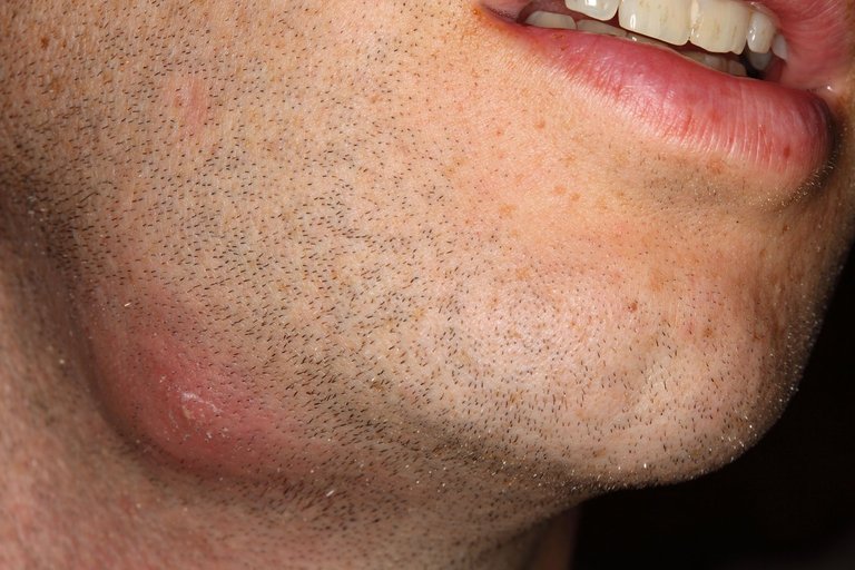
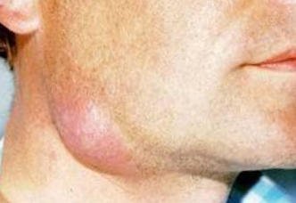
- Infecciones del espacio submandibular: son producidas más frecuentemente por causas odontogénicas, infección de glándulas salivales (submandibular y sublingual).
Clínica: dolor, fiebre, aumento de volumen en región submandibular con signos de flogosis.
Infecciones del piso de la boca: la odinofagia es el síntoma más importante, glosodinia, protrusión lingual hacia arriba y hacia atrás, trismo, aumento de volumen submandibular, dificultad respiratoria en casos severos, fiebre. Entre las infecciones del piso de boca existe una forma clínica que cursa con celulitis, denominada angina de Ludwig, enfermedad grave que puede poner en serio compromiso la función ventilatoria del paciente.
Fascitis necrotizante: puede originarse en cualquier espacio cervical, de rápida evolución hacia otros espacios. Se caracteriza por dolor intenso, edema con parches de tejido necrótico, enfisema subcutáneo y deterioro progresivo del estado general.
Peritonsillar space infection: Associated with pharyngotonsillar complications.
Infection of the retropharyngeal space: differs in children and adults due to the presence of lymphatic tissue. The most frequent etiologies in children are upper respiratory infections (acute and chronic adenoiditis); in adults, foreign bodies and surgical procedures or associated diseases.
Clinical: increased volume of posterior wall of rhinopharynx and oropharynx, torticollis, inspiratory stridor, sialorrhea, closed rhinolalia, intense odynophagia, progressive dysphagia, fever.
Infection of the parapharyngeal space: for better systematization, it should be individualized in the pre-stylar and retro-stylar space.
Pre-stylar: dissemination of tonsillar infections, local iatrogenic maneuvers, parotiditis and intraparotid lymphadenitis.
Clinical: odynophagia and severe dysphagia associated with reflex otalgia, sialorrhea, trismus, fever, torticollis, lateral cervical enlargement, lateropharyngeal bulges that are located behind the tonsil and push it forward and towards the midline.
- Retroesthesia: these are infections of greater severity because they contain the internal and external carotid arteries, the internal jugular vein, the superior cervical sympathetic and cranial nerves IX, X, XI, XII. It can be of tonsillar, odontogenic and rhinosinusal origin.
Clinical symptoms: general condition, high fever, torticollis, pain on cervical palpation, and pharyngeal symptoms are scarce with laterocervical enlargement and signs of phlogosis.
- Infections of the submandibular space: they are most frequently produced by odontogenic causes, infection of salivary glands (submandibular and sublingual).
Clinical: pain, fever, increase of volume in the submandibular region with signs of phlogosis.
Mouth floor infections: odynophagia is the most important symptom, glossodynia, upward and backward tongue protrusion, trismus, submandibular enlargement, respiratory distress in severe cases, fever. Among the infections of the floor of the mouth there is a clinical form with cellulitis, called Ludwig's angina, a severe disease that can seriously compromise the patient's ventilatory function.
Necrotizing fasciitis: it may originate in any cervical space, with rapid evolution towards other spaces. It is characterized by intense pain, edema with patches of necrotic tissue, subcutaneous emphysema and progressive deterioration of the general condition.

DIAGNÓSTICO
DIAGNOSTIC
Examen físico de cabeza y cuello, con ubicación de los espacios anatómicos comprometidos. La evaluación de la vía aérea con endoscopio rígido o flexible debe ser realizada por el otorrinolaringólogo.
Laboratorio: hematología completa, reactantes de fase aguda (VSG, Proteína C reactiva, procalcitonina), ácido láctico, cultivos de secreción y tejidos, serología para hongos. La elevación de la VSG, la leucocitosis moderada a severa, la trombocitosis y la relación N:L superior a 1,6 representan parámetros de laboratorio al momento del ingreso, que permiten prever la aparición de complicaciones y la importancia de realizar controles sucesivos durante el periodo crítico de las primeras 48 horas y la decisión de realizar algún procedimiento quirúrgico.
Estudios de imágenes: se ha demostrado la utilidad de la tomografía computada y la resonancia magnética con contraste en el diagnóstico clínico de las IPC, antes de decidir la conducta terapéutica.
Physical examination of the head and neck, with location of the anatomical spaces involved. Evaluation of the airway with rigid or flexible endoscope should be performed by the otolaryngologist.
Laboratory: complete hematology, acute phase reactants (ESR, C-reactive protein, procalcitonin), lactic acid, secretion and tissue cultures, fungal serology. Elevated ESR, moderate to severe leukocytosis, thrombocytosis and N:L ratio higher than 1.6 represent laboratory parameters at the time of admission, which allow predicting the appearance of complications and the importance of successive controls during the critical period of the first 48 hours and the decision to perform a surgical procedure.
Imaging studies: the usefulness of computed tomography and magnetic resonance imaging with contrast has been demonstrated in the clinical diagnosis of CPI, before deciding on the therapeutic approach.

De acuerdo a la IPC y sus probables complicaciones se dispone de:
According to the IPC and its probable complications, the following are available
• Radiografía lateral de cuello
• Panorámica dental (en sospecha de infección odontogénica)
• Radiografía de tórax PA y lateral
• Ultrasonido
• Tomografía computada de cuello con contraste, resonancia magnética.
Lateral neck x-ray
Dental panoramic view (in suspicion of odontogenic infection).
PA and lateral chest radiographs
Ultrasound
Computed tomography of the neck with contrast, magnetic resonance imaging.

El tratamiento quirúrgico se debe considerar en las siguientes indicaciones:
Surgical treatment should be considered in the following indications:
• Compromiso de la vía aérea
• Condiciones críticas
• Septicemia
• Sospecha de fascitis necrotizante por presencia de gas o niveles hidroaéreos
• Falta de respuesta a antibióticos empíricos después de 48 horas o persistencia posterior a punción simple
• Infecciones descendentes
• Colecciones multiloculares o con compromiso de dos o mas espacios
• Abscesos mayores de 3 cm de diámetro ubicados en los espacios visceral anterior, submandibular, parafaringeo, prevertebral, carotideo y retrofaríngeo
• Cuenta blanca mayor de 20.700
• Abscesos con diámetro mayor de 22 mm en tomografía
• Pacientes menores de 51 meses de edad que requieran terapia intensiva
Airway compromise
Critical conditions
Sepsis
Suspicion of necrotizing fasciitis due to the presence of gas or hydroaerial levels.
Failure to respond to empirical antibiotics after 48 hours or persistence after simple puncture
Descending infections
Multilocular collections or with involvement of two or more spaces
Abscesses greater than 3 cm in diameter located in the anterior visceral, submandibular, parapharyngeal, prevertebral, carotid and retropharyngeal spaces.
White count greater than 20,700
Abscesses with a diameter greater than 22 mm in tomography.
Patients less than 51 months of age requiring intensive therapy.

Complicaciones:
Complications:
1.- Obstrucción de la vía aérea
2.- Broncoaspiración
3.- Complicaciones vasculares: trombosis de la vena yugular interna, ruptura de la arteria carótida
4.- Mediastinitis como extensión del proceso infeccioso
5.- Fascitis necrosante del cuello
6.- Déficit neurológico por compromiso de los pares craneales
7.- Embolia séptica a nivel pulmonar, cerebral y articulaciones
8.- Osteomielitis de estructuras óseas de la base del cráneo, mandíbula y columna cervical.
9.- Síndrome de Grisel (tortícolis inflamatorio causado por sub-luxación de vértebras cervicales).
10.- Shock séptico; la mediastinitis descendente y la obstrucción de las vías respiratorias son las complicaciones más temidas de la IPC. En casos con afectación del mediastino superior es necesario la mediastinotomía transcervical. Esto tiene una alta tasa de morbilidad y mortalidad.
1.- Airway obstruction
Bronchoaspiration
3.- Vascular complications: thrombosis of the internal jugular vein, rupture of the carotid artery.
4 .- Mediastinitis as an extension of the infectious process
5.- Necrotizing fasciitis of the neck.
Neurological deficits due to cranial nerve involvement.
7.- Septic embolism at pulmonary, cerebral and joint level.
Osteomyelitis of bony structures of the base of the skull, jaw and cervical spine.
Grisel's syndrome (inflammatory torticollis caused by sub-luxation of cervical vertebrae).
Septic shock; descending mediastinitis and airway obstruction are the most feared complications of CPI. In cases with involvement of the superior mediastinum, transcervical mediastinotomy is necessary. This has a high morbidity and mortality rate.

Bueno amigos, espero les haya gustado esta información, como se podrán dar cuenta, las infecciones profundas de cuello, atentan contra la vida, dado que, pueden ocupar con pus las vías aéreas y fallecer por asfixia mecánica, o, puede que hagan una sepsis general y morir por bacterias en todo el organismo, por favor, tomemos conciencia, y eliminemos todos los focos de caries que tengamos en nuestra boca, porque por allí comienza esta patología y cuando poco, están comprometidos e incluso fallecer, así que pilas, visiten al odontólogo y a su médico especialista el Otorrinolaringólogo, su mejor aliado. Feliz Día...
Well friends, I hope you liked this information, as you can see, deep neck infections are life threatening, because they can occupy the airways with pus and die from mechanical asphyxia, or may cause a general sepsis and die from bacteria throughout the body, please, let's be aware and eliminate all the caries that we have in our mouth, because that is where this pathology begins and when little, they are compromised and even die, so batteries, visit the dentist and your specialist doctor, the Otolaryngologist, your best ally. Happy day...

√ ABSCESO PROFUNDO DE CUELLO HOSPITAL JUAREZ
√ PATOLOGÍA INFLAMATORIA CERVICAL


Electronic-terrorism, voice to skull and neuro monitoring on Hive and Steem. You can ignore this, but your going to wish you didnt soon. This is happening whether you believe it or not. https://ecency.com/fyrstikken/@fairandbalanced/i-am-the-only-motherfucker-on-the-internet-pointing-to-a-direct-source-for-voice-to-skull-electronic-terrorism
https://twitter.com/JosCarrizalez3/status/1448835737697259521
The rewards earned on this comment will go directly to the person sharing the post on Twitter as long as they are registered with @poshtoken. Sign up at https://hiveposh.com.
Congratulations @krrizjos18! You have completed the following achievement on the Hive blockchain and have been rewarded with new badge(s) :
Your next target is to reach 10000 upvotes.
You can view your badges on your board and compare yourself to others in the Ranking
If you no longer want to receive notifications, reply to this comment with the word
STOPExcelente post 👏👏👏
muchas gracias.... espero les sirva de mucho provecho....
Tu , publicación es muy interesante..una enfermedad que se puede prevenir...
si señor en efecto.... se puede prevenir siempre y cuando tomemos conciencia y atendamos nuestras caries q por lo general es el factor primordial....